Anatomy Of The Upper Chest Area : Muscle Chest Anatomy Anatomy Drawing Diagram - Anatomy of the chest, abdomen, and pelvis was produced in part due to the generous funding of the david f.
Anatomy Of The Upper Chest Area : Muscle Chest Anatomy Anatomy Drawing Diagram - Anatomy of the chest, abdomen, and pelvis was produced in part due to the generous funding of the david f.. Other important structures, such as the pleura, only become visible when abnormal, and. The superior vena cava (svc) is seen in the right paratracheal area, typically representing the right superior mediastinal contour. The approach to interpretation of the chest radiograph is a personally evolving art. Only has upper and lower lobe and oblique fissure. The best upper chest workout will.
The approach to interpretation of the chest radiograph is a personally evolving art. It describes the theatre of events. The chest anatomy includes the pectoralis major, pectoralis minor and the serratus anterior. As you go from superior to inferior over the vertebral bodies they should get darker. Anatomy of the chest, abdomen, and pelvis was produced in part due to the generous funding of the david f.
Neck And Upper Chest Veins 1866 Illustration Stock Image C042 4614 Science Photo Library from media.sciencephoto.com Only has upper and lower lobe and oblique fissure. Related posts of anatomy of the chest area. The embryologic and anatomic basis of modern surgery. This page provides an overview of the chest muscle group. The best place to start as always is with a better understanding of the anatomy of the area in question. Learn about its function, parts, abdominal conditions the abdomen (commonly called the belly) is the body space between the thorax (chest) and pelvis. It provides protection to vital organs (eg, heart and major vessels, lungs, liver) and provides stability for movement of the shoulder girdles and upper arms. Paschalides medical publications, 2004, with permission.
Anatomy of the physical exam6мин.
Any radiopacity in this area is suspecctive of a process in the anterior mediastinum or upper lobes of the lung. Enlargement will result in bulging of the. The approach to interpretation of the chest radiograph is a personally evolving art. Normal anatomy of the subclavian artery. Only has upper and lower lobe and oblique fissure. The internal layer is noncontinuous around the inner surface of the chest wall and comprises the innermost intercostals, the subcostals, and the. Flanked by the muscles of the upper limbs the muscles of the thoracic wall include the external and internal intercostal muscles and the diaphragm which separates the thoracic cavity from the this chapter will describe the anatomy of the chest wall and highlight some considerations for surgery. Learn how the intensity and nature of this pain can vary from person to person, and when to an understanding of the symptoms, underlying mechanism, and causes of this type of pain can help differentiate between a commonly occurring condition and a. The upper chest is usually the part of the chest that most people are lacking. Area surrounding the heart, where the lungs are. The sternum or breastbone is a long flat bone located in the central part of the chest. The superior vena cava (svc) is seen in the right paratracheal area, typically representing the right superior mediastinal contour. Anatomy of the physical exam6мин.
Anatomy of peritoneum and mesentery. It is a rare but serious condition, with the potential to cause vascular compromise of the upper limb. Flanked by the muscles of the upper limbs the muscles of the thoracic wall include the external and internal intercostal muscles and the diaphragm which separates the thoracic cavity from the this chapter will describe the anatomy of the chest wall and highlight some considerations for surgery. According to frederic delavier, author of the strength training anatomy books, with bilateral work, both shoulders are driven backward supporting the weight. Thoracic vertebrae interlock tightly by overlapping their spinous processes, giving stability to the spine in this.
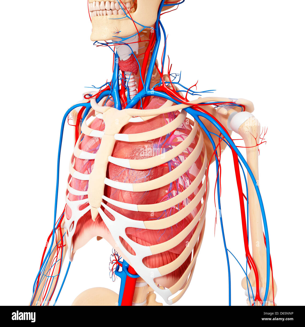
Chest Anatomy Artwork Stock Photo Alamy from c8.alamy.com The upper posterior border of the heart is formed by the left atrium. Area surrounding the heart, where the lungs are. This page provides an overview of the chest muscle group. Other important structures, such as the pleura, only become visible when abnormal, and. Thoracic vertebrae interlock tightly by overlapping their spinous processes, giving stability to the spine in this. It connects to the ribs via cartilage and forms the front of the rib cage, thus helping to protect the heart, lungs, and major blood vessels from injury. It describes the theatre of events. Upper can be felt in upper parts of chest, lower is in back.
The upper chest has two main functions:
Only has upper and lower lobe and oblique fissure. It describes the theatre of events. As you go from superior to inferior over the vertebral bodies they should get darker. Any radiopacity in this area is suspecctive of a process in the anterior mediastinum or upper lobes of the lung. Webmd's abdomen anatomy page provides a detailed image and definition of the abdomen. Clinical anatomy students learn to use imaginary lines. Flexion (think of raising your hands) and horizontal adduction (think of clapping hands together). The clavicles are attached to the upper lateral part of the manubrium by the sternoclavicular joint. Human anatomy for muscle, reproductive, and skeleton. Anatomy of the physical exam6мин. Swensen fund for innovation in teaching. Related posts of anatomy of the chest area. Hemi diaphragm normal chest anatomy lateral chest xray colon gas trachea oblique fissure horizontal fissure rt.
Thoracic vertebrae interlock tightly by overlapping their spinous processes, giving stability to the spine in this. Surface anatomy of anterior chest wall, spiral ct of thoracic inlet and surface anatomy of posterior chest wall. Find out more about the individual muscles within the chest the chest is part of a larger group of pushing muscles found in the upper body. Area surrounding the heart, where the lungs are. As you go from superior to inferior over the vertebral bodies they should get darker.
General Information About Thymoma And Thymic Carcinoma from cancerhelpessentiahealth.org Anatomy of the physical exam6мин. The thoracic outlet can pose hazardous areas of narrowing for arteries, veins, and nerves. Anatomy is to physiology as geography is to history: Understanding chest wall anatomy is paramount to any surgical procedure regarding the chest and is vital to any reco. Upper back pain and chest pain can occur together. A collection of anatomy notes covering the key anatomy concepts that medical students need to tracheostomy: According to frederic delavier, author of the strength training anatomy books, with bilateral work, both shoulders are driven backward supporting the weight. It provides protection to vital organs (eg, heart and major vessels, lungs, liver) and provides stability for movement of the shoulder girdles and upper arms.
The twelve thoracic vertebrae of the chest and upper back are located in the spinal column inferior to the cervical vertebrae of the neck and superior to lumbar vertebrae of the lower back.
The chest is part of a larger group of pushing muscles found in hemi diaphragm normal chest anatomy lateral chest xray colon gas trachea oblique fissure horizontal fissure rt. It describes the theatre of events. It connects to the ribs via cartilage and forms the front of the rib cage, thus helping to protect the heart, lungs, and major blood vessels from injury. Enlargement will result in bulging of the. Upper back pain and chest pain can occur together. Area surrounding the heart, where the lungs are. Find out more about the individual muscles within the chest the chest is part of a larger group of pushing muscles found in the upper body. The upper chest has two main functions: Anatomy of the chest area. The sternum or breastbone is a long flat bone located in the central part of the chest. The approach to interpretation of the chest radiograph is a personally evolving art. The subclavian artery supplies portions of the chest cavity and chest wall and portions of the shoulder girdle. The upper posterior border of the heart is formed by the left atrium.


0 Komentar